

Table of Contents
- Understanding Cranio-Cervical Instability
- Limitations of Traditional MRI in CCI Diagnosis
- The Role of Dynamic MRI Imaging
- Integration of Artificial Intelligence in MRI
- Emergence of Wearable MRI Technologies
- Ultra-Low Field MRI: A New Frontier
- Conclusion
Magnetic Resonance Imaging (MRI) remains a vital tool for evaluating spinal and neurological conditions. The last decade has seen remarkable progress in imaging technology, most notably in addressing the unique challenges related to diagnosing cranio-cervical instability (CCI). These technological advancements now enable specialists to obtain clearer, more dynamic views of the joint between the skull and the upper cervical spine, leading to more reliable diagnoses and better patient outcomes. For individuals facing persistent symptoms like unexplained headaches, neck pain, or neurological deficits, getting an accurate cervical spine MRI can be instrumental in uncovering the causes and determining the right treatment plan.
Cranio-cervical instability (CCI) presents variably among patients, often becoming evident only under specific physical stresses. Advances in imaging technology, particularly MRI, enable medical professionals to observe subtle abnormalities in real time by evaluating patients in different physiological positions, seated, upright, or during motion. This method allows clinicians to effectively assess changes in stability with posture. The National Institutes of Health (NIH) and other health organizations emphasize the importance of advanced imaging for accurately diagnosing conditions like CCI, a complex spinal disorder characterized by abnormal motion or excess laxity between the skull and the cervical spine. Traditional diagnostic methods have faced challenges due to vague symptoms and limitations of conventional imaging. However, modern MRI capabilities provide powerful tools for visualizing the dynamic response of these joints during movement. The integration of detailed anatomical imaging with functional assessment marks a significant advancement in recognizing and managing CCI, leading to more sophisticated, patient-centered diagnostic protocols.
Understanding Cranio-Cervical Instability
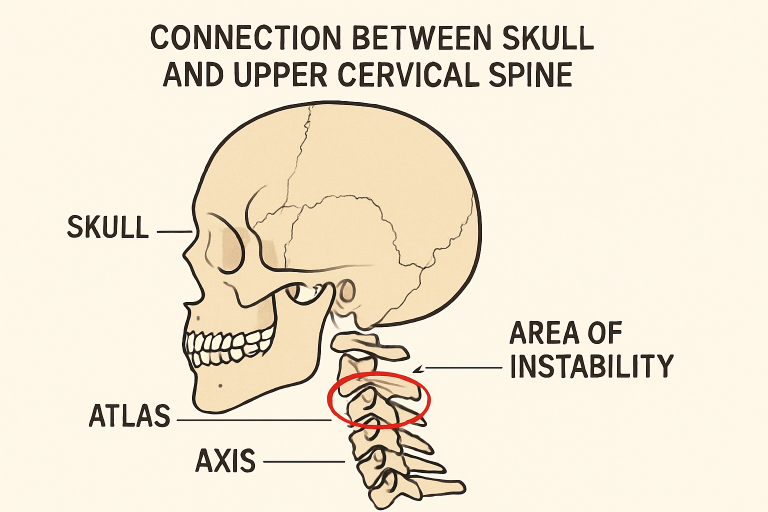
CCI primarily refers to abnormal movement at the junction of the skull (cranium) and the upper cervical spine, particularly involving the atlas (C1) and axis (C2) vertebrae. Individuals with CCI may experience a broad spectrum of symptoms, including severe neck pain, headaches, dizziness, blurred vision, and neurological deficits like weakness or tingling in the limbs. The causes range from connective tissue disorders, such as Ehlers-Danlos Syndrome, to trauma or inflammatory diseases.
Accurately diagnosing CCI is essential because, without proper identification and management, continued instability can lead to significant neurological complications. Therefore, imaging has become an integral part of identifying the underlying causes and planning effective treatment strategies.
Limitations of Traditional MRI in CCI Diagnosis
Traditional MRI systems have served clinicians for decades, but their standard design, which requires patients to lie flat and remain still, may fall short when identifying conditions like CCI. Because these scans only capture static, supine images, dynamic or position-dependent instabilities often go undetected. For CCI, the full extent of instability may only become evident when the neck is subjected to specific types of physical stress, which cannot be replicated in a standard MRI scanner.
This limitation can lead to misdiagnoses or missed diagnoses, causing patients to experience prolonged suffering and delayed interventions. As a result, the demand for more versatile imaging solutions has driven the industry to innovate beyond traditional approaches.
The Role of Dynamic MRI Imaging
Dynamic MRI, also known as motion or upright MRI, captures images while the patient moves their head and neck through various positions. By doing so, radiologists and clinicians can observe real-time shifts in alignment and joint stability that would not be apparent in static images. This imaging method is particularly valuable for assessing patients with suspected CCI due to connective tissue disorders or previous cervical trauma.
A landmark study of individuals with Ehlers-Danlos Syndrome demonstrated that dynamic MRI detected instability that conventional imaging missed. This aligns with clinical observations that position-dependent CCI may only manifest on positional imaging.
Integration of Artificial Intelligence in MRI
Artificial intelligence has emerged as a game-changer in the analysis of MRI scans. AI-driven software can automate the identification of cervical spine abnormalities and distinguish between subtle changes in vertebral position, ligament laxity, or abnormal joint motion. These systems leverage large datasets to train neural networks, enabling faster, more accurate assessment of complex imaging studies.
When integrated into MRI diagnostics, AI reduces human error, shortens image processing time, and increases precision, especially in cases of cervical spondylosis and CCI. Several clinical trials have reported that AI-assisted interpretation correlates strongly with expert radiologist review.
Emergence of Wearable MRI Technologies
Wearable MRI devices are another breakthrough in medical imaging. Using lightweight, flexible metamaterials that patients can wear, these devices offer more comfortable and accessible imaging experiences. They enable clinicians to acquire images in natural conditions, without the constraints of traditional MRI systems. Early research suggests that wearable MRIs improve signal-to-noise ratio, resulting in clearer, more detailed images.
These innovations make it easier for those with mobility challenges or severe pain to undergo imaging, and they hold promise for expanding the reach of advanced diagnostics to outpatient clinics and remote care settings.
Ultra-Low Field MRI: A New Frontier
Ultra-Low Field MRI (ULF MRI) systems represent another significant step forward in imaging technology. Designed for portability and cost-effectiveness, ULF MRI employs magnets well below the strength of conventional scanners. Despite lower field strengths, these devices can visualize white matter microstructure, nerve pathways, and subtle changes relevant to cranio-cervical junction disorders.
Recent studies have demonstrated reliable tractography with ULF MRI, a form of imaging that maps nerve fiber connections within the spinal cord and brainstem. This offers a new approach for clinicians to detect neurological implications of instability, particularly in patients who may not tolerate traditional MRI setups or require long-term monitoring.
Conclusion
The ongoing evolution of MRI technology is reshaping how clinicians diagnose and treat cranio-cervical instability. From dynamic and wearable imaging to the integration of artificial intelligence and portable low-field devices, patients and providers now have a suite of effective diagnostic options. These advancements not only improve diagnostic accuracy but also enhance comfort, accessibility, and overall patient care. As the field advances, patients with persistent neck or neurological symptoms should consider advanced imaging as an essential part of their diagnostic process to improve outcomes and quality of life.

FOLLOW ME ON SOCIAL MEDIA


No comments:
Post a Comment
I love reading and responding to comments but in order to get my reply you must ensure you are NOT a no-reply blogger. If you are, here are some quick steps to change that!
1. Go to the home page of your Blogger account.
2. Select the drop down beside your name on the top right corner and choose Blogger Profile.
3. Select Edit Profile at the top right.
4. Select the Show My Email Address box.
5. Hit Save Profile.